

Study Participants and Volunteers

Welcome to the University of Delaware’s Center for Biomedical and Brain Imaging (CBBI). This state-of-the-art research facility houses the first research fMRI scanner in the state of Delaware—with the capability to map brain activity, as well as provide remarkably detailed images of muscles, discs, bones and organs. Serving researchers campuswide, statewide and throughout the region, the CBBI will advance research on psychopathology, cancer, stroke, cerebral palsy, osteoporosis and other diseases and disorders.

Research Partnerships
CBBI is actively working to build long-term partnerships with campus and regional researchers. Information on partnership opportunities will be coming soon.
UD Research
UD’s Center for Biomedical and Brain Imaging, an 11,600-square-foot, two-story extension to the Life Sciences Research Facility, houses the state’s first research dedicated MRI scanner, which can be used to measure brain structure and function. Functional MRI (fMRI) shows the function of different brain areas responsible for various mental activities such as feeling, emotion, retrieving a memory, etc. and does so in a safe, non-invasive way. The instrument, which is located on the first floor, also can also provide ultra-high-resolution images of muscles and skeletal structures in humans and animals. The second floor includes a conference/teaching area and bench-top imaging instruments.


CBBI Capabilities
Our Siemens Magnetom Prisma whole-body MRI scanner, with its 3 Tesla magnet, provides stunningly detailed images, allowing researchers in multiple disciplines to examine the internal structures of living beings in a safe, non-invasive way. The 3T suite is equipped with visual and auditory stimulation equipment, eye-tracking, response devices and much more. Space has been allocated for future expansion.
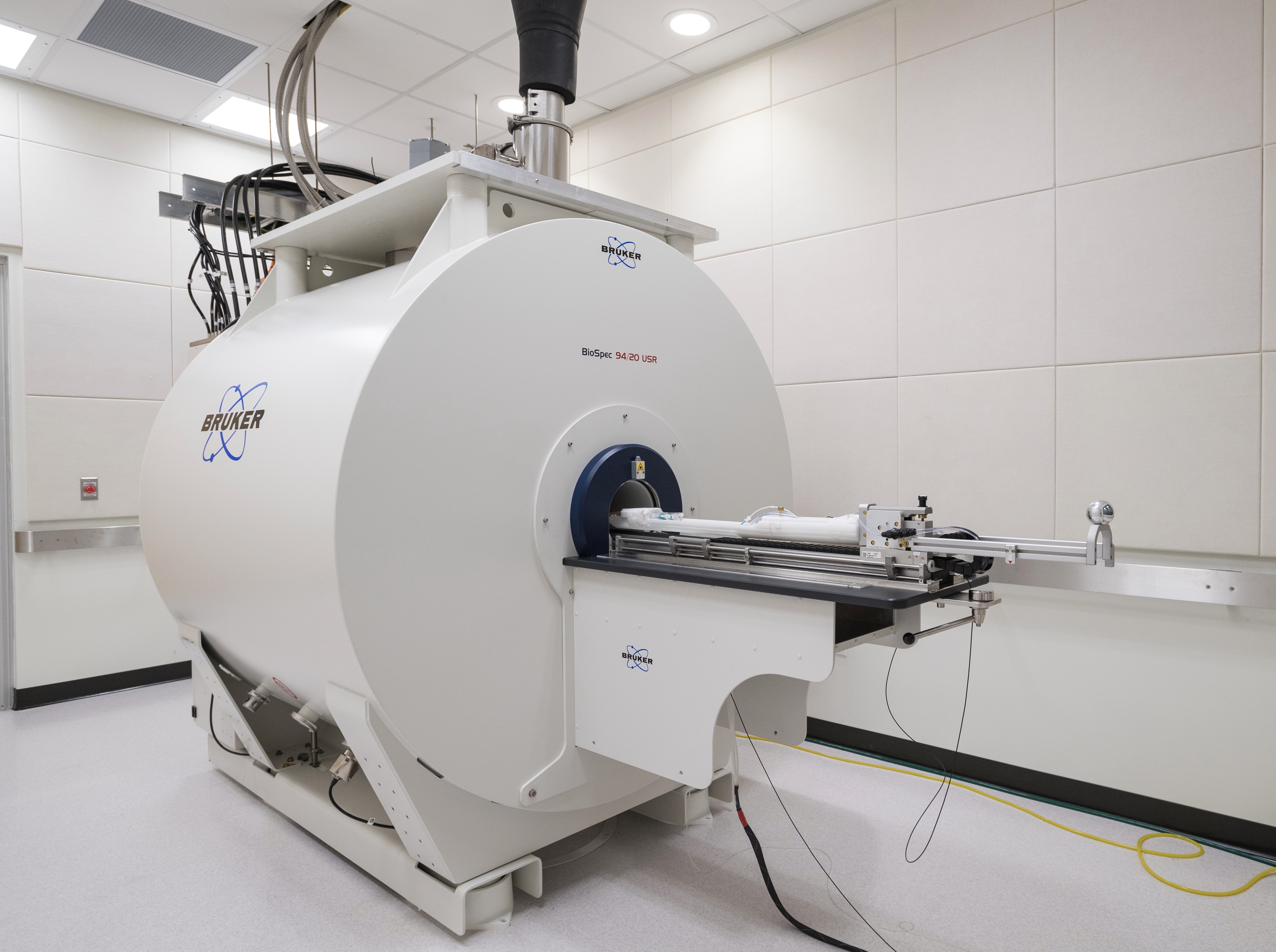
Our Bruker Biospec 94/20, a 9.4 Tesla magnet, designed for pre-clinical MR imaging and MRI research. State-of-the-art MRI technology combined with the ultra high field magnetic field delivers high spatial resolution, enabling researchers to come closer to the molecular and cellular level research they desire.
How does an fMRI work?
The “f” in the fMRI refers to “function.” This special kind of MRI will help scientists understand what areas of the brain are most active when a person performs a mental task. fMRI reveals which parts of the brain are stimulated, or “at work,” by measuring the blood flow.
What is fMRI used for?
The fMRI technique can reveal the brain regions that are active when a study participant is engaged in cognitive activities. It can shed light on myriad topics, from tumor development, to damage to discs in the spine, to the structure of next-generation materials and much more.
Is the imaging harmful?
Imaging by fMRI poses almost no risk to the average subject when appropriate safety guidelines are followed. It is non-invasive and uses radio waves (no X-rays). Although its strong magnetic field is not harmful in itself, implanted medical devices that contain metal may be affected.
Trevor Wigal
Manager, CBBI
Phone: 302-831-1463
Xingju Nie
MRI Physicist
Phone: 302-831-0648
Ibrahim Malik
Research Associate
Phone: 302-831-0910
Sarah Whitman
Research Associate
Phone: 302-831-0910
HOURS OF OPERATION
The University of Delaware Research Office is open daily Monday through Friday, 8 a.m.– 5 p.m. The Research Office is closed Saturday and Sunday and observes all University closings and holidays. See the Academic Calendar for more information.
If you have an emergency during non-business hours, please call Public Safety at 302-831-2222.
If your question does not require immediate attention, please e-mail CBBI.