

Safety Training
The static magnetic field of an MRI system is always present. It is important that all those entering the facility are properly trained and aware of the presence of these fields.

Research involving Magnetic Resonance Imaging (MRI) at high magnetic field strengths presents unique challenges to both research participants and individuals working within and around the MRI system.
MRI Facility Personnel
Level 1 MR Personnel
Definition: Level 1 MR Personnel are Individuals who have had MRI safety training as approved by the CBBI manager and/or director.
Facility access allowance: Upon completion of Level 1 training, Level 1 MR Personnel are permitted to be in Zones I-III of the CBBI. Level 1 users are permitted in Zone IV under the supervision of a Level 2 user.
Restrictions: Level 1 users may not operate the scanner or enter Zone IV without supervision or administer the MRI screening to others.
Documentation of Level 1 MR Personnel qualification: Documentation of Level 1 MR Personnel qualification is maintained within the CBBI.
Level 2 MR Personnel
Definition: Level 2 MR Personnel have completed all requisite MRI safety and user training established by the CBBI. Level 2 users may operate the scanner without supervision, supervise Level 1 users, and administer the MRI safety screening to study participants.
Facility Access Allowance: Level 2 users may access Zones I-IV without supervision.
Documentation of Level 2 MR Personnel qualification: Records are maintained within the CBBI.
Additional requirements
A minimum of two persons are required at all times when operating the scanner, one of which must be a Certified Level 2 User.
RISK OF INJURY
- Static Magnetic Field Risk. The magnetic field of a MRI machine is exceptionally strong. The field attracts certain metals and draws them to the magnet with uncontrollable force. This may damage the magnet or strike and injure a patient or employee near the magnet. Ferromagnetic metals (most commonly iron and some forms of steel) are most strongly attracted to magnets. Some other metals, such as cobalt, nickel, titanium, chromium, platinum and certain alloys, may be attracted to a much lesser degree. Examples of objects that may become lodged in the magnet include gurneys and beds (especially those with motors), Intravenous Therapy (IV) poles, oxygen tanks, and floor buffers. Implanted spring steel, such as early generation intracranial aneurysm clips, may become dislodged or twist in the magnetic field causing an intracranial hemorrhage. Steel fragments near the orbit may injure the optic nerve. Pacemakers and other medical devices may be damaged, reprogrammed, or turned off.
- Radio-frequency (RF) Electromagnetic Field Risk. Prolonged imaging may cause the patient’s core body temperature to rise by deposition of energy from RF fields. The RF field may induce currents in electrically-conductive materials, such as wires that are lying on the patient, causing skin burns. It may also induce currents in intra-cardiac leads, resulting in inadvertent cardiac pacing.
- Gradient Magnetic Field Risk. While changes in magnetic fields do not cause harm under normal imaging conditions, extreme gradients may cause biological effects, such as contraction of peripheral muscles or perceived flashes of light from the retina. Potentially, extreme gradients could induce seizures or arrhythmias.
- Cryogen Risk. During an unplanned loss of magnetic field, helium in the magnet may evaporate suddenly. This gas is normally directed to the exterior of the building by large pipes. In the event that helium leaks into the magnet room, it may displace oxygen in the room which could result in suffocation.
BMC Medical Imaging 2013 13:11 DOI: 10.1186/1471-2342-13-11
© Tokue et al.; licensee BioMed Central Ltd. 2013
MAGNETIC RESONANCE IMAGING HAZARDS
Hazards to personnel and patients in the MRI suite can be categorized as follows:
TRANSLATIONAL FORCES—
THE MISSILE EFFECT
This effect is generally attendant upon ferromagnetic materials and the static field generated by an MRI system, and often manifests as the missile effect, which can involve non-compatible objects, such as wheel chairs, transport stretchers, file cabinets, electrical equipment and tools, powered and unpowered hand tools, stethoscopes, medical equipment carts, medical equipment (pulse oximeters, EKG, IV pumps, etc), communications devices, physician beepers, and miscellaneous (and often forgotten) patient and visitor objects that include hairpins, paper clips and pens.
A hairpin or paper clip within the 5-10 gauss line range could reach a velocity of 40 mph and will be attracted to the center of the MRI generated field (x, y and z axis) where the lines of force are equal.
TORQUE FORCES
Torque forces run parallel to the translational force. Both are associated with ferromagnetic materials and the static field generated by an MRI machine. Attracted objects react by aligning parallel to the magnetic lines of force being generated by the MRI machine. The center of the MRI-generated field has the highest torque force, creating a serious exposure for all contraindicated items and MRI-conditional items in the MRI suite, depending on the tesla rating of the MRI. This effect has created life-threatening conditions for patients with some medical implants.
INDUCED MAGNETIC FIELDS
Any metallic object introduced into a high flux field will induce a current if that object is moving and perpendicular to the force lines. That new induced current will create a secondary magnetic field that will oppose the original field.
The same metallic object introduced parallel to the force lines will generate no effect. While not as apparent as translational forces, induced magnetic fields can cause patients discomfort or anxiety due to reactive forces on MRI-safe medical implants.
All pacemakers and implantable cardioverter/defibrillators should be considered contraindicated under any circumstance. When these devices are exposed to an MRI environment, a life-threatening condition may be created within the five-gauss line. Intravascular catheters, intubation equipment, infusion pumps, orthopedic implants, stents and other devices should be verified as MRI-safe in the prescreening. The transportation of a patient should be designed to minimize potential torque from secondary magnetic fields by maintaining a parallel path to the lines of force. Any MRI-safe device must be verified as acceptable for the MRI tesla-rated machine and maximum closest safe distance from the machine.
THERMAL HEATING
The static magnetic MRI field will tend to induce currents in any conductive materials, including those that may be non-ferrous. However, the human body is electrically conductive by nature and small RF fields will generate current that will be absorbed by the body as heat. The specific absorption rate (SAR) for the patient will depend on weight and body radius. The heating will be more prominent at the periphery of the body than at its core. This effect will also vary with the type of MRI scanning being done (angle, pulse frequency, fast spin echo, etc.).
The most serious exposure is located in the bore of the MRI machine and in the axis points, as they possess the highest potential torque forces. The use of extremity coils could aggravate the risk, but can be avoided through the use of MRI-safe polymeric foam padding. In addition, monitoring patient position in the MRI machine and proximity to its inner wall should be included in patient care protocol.
The most common sources of thermal exposure tend to be looped/un-looped medical equipment leads, MRI accessories, and sensors. These include conductive loops touching the patient or crossing the extremities, clothing, and metalized drug delivery patches (OTC or prescription).
PATIENT DISCOMFORT
Secondary induced currents and subsequent secondary generated magnetic fields may cause brief and benign – but noticeable – reactions in the patient.
Closed bore MRI machines can cause a patient discomfort if the patient is claustrophobic to any degree.
Loud noises, light flashes and other sensory input may be perceived by the patient.
Neonates (premature infants) and elderly patients may be at higher risk due to changes in ventilation and hemodynamic stability, and vasoactive infusion requirements. Close communication and cooperation between MRI staff and the attending anesthesiologist is critical.
QUENCHING
MRI machines are cooled by a super-cooling fluid (liquid helium). The release of the super-cooling fluid into the atmosphere is called quenching, and unintentional and intended magnet quenching can be catastrophic. Most clinical machines have a 700 to 1,000 liter-volume of this cryogenic. Its venting will cause oxygen in the MRI magnet room to condense around the vent pipe and accumulate in the MRI machine, posing a code red fire hazard.
Quenching is normally associated with de-energizing the machine, which is safer then a quenching during a scanning procedure. However, the room will still be subject to increased levels of oxygen near the machine. Not only should the patient be removed as quickly as possible during the de-energizing process, but any sources of possible ignition near the machine should be minimized. Room exhaust ventilation should be activated. Another risk is a quench vent pipe breech, which could flood the room with cryogenic fluids. This creates an asphyxiation hazard for the patient and attending staff.
If an emergency should arise in the MRI machine room, all staff and respondents must be aware of the potential risks of translational and torque induced effects on the patient. All code blue equipment must be located outside the five-gauss line to maintain an MRI-safe environment. Both code red and code blue situations will require preplanning with hospital medical staff and emergency response teams. Municipal firefighters should not be allowed into the room until the MRI is proven to be de-energized or they are MRI-safe – with magnetic equipment and clothing removed.
MR Safety Guide
General Rules for the MR Suite
-
- Before a research study partcipant enters the magnet room, a screening form must be completed by the subject and reviewed by the certified user who will be performing the MRI exam.
- Any person not receiving an MRI as part of a research study or any person not previously screened and trained in the CBBI procedures may not enter the MRI room without approval from CBBI staff. (maintenance workers, custodial staff, etc.)
- Before entering the magnet room, all pockets are to be emptied of: watches, pagers, wallets, pens, pencils, hair clips, jewelry, keys, coins, and any other possible projectiles.
- The person being scanned must be wearing MRI safe attire.
Scrub tops and bottoms are provided for this purpose.
-
- The magnet room door should be kept closed at all times except when entering or exiting the room.
Keeping the door shut tightly will reduce RF noise during imaging.
- All persons scanned must wear ear protection. I f they decline, the exam must be canceled.
- Never scan with an unplugged surface (or body flex) coil in the bore of the magnet.
- Use of any research equipment, coils, or supplies not supplied by the MRI facility must be approved by an MR Safety Committee or appropriate official.
- All investigators and operators must clean up after themselves. Return all equipment to its proper place. The room should be kept neat and tidy. Do not lay coils or phantoms on the floors.
- Please report all malfunctioning or damaged equipment to the center manager immediately.
- All studies should be archived immediately after the exam is complete. Data may remain on the system for up to 7 days (or some agreed upon time) unless disk space becomes critical, in which case, the oldest studies will be removed first.
- All users must be familiar with code procedures and calling 911 if a medical emergency should arise.
- Users must never leave their subjects unattended in the magnet.
Section I: The Magnetic Environment
Magnetic Field
It is important to remember when working around a superconducting magnet that the magnetic field is always on. Under normal working conditions, the field is never turned off. Therefore, it is important to be aware of safety issues regarding ferrous objects that can act as projectiles and watching out for patients who may have contraindicated devices implanted in their bodies. There are two units used to describe magnetic field strength. They are Tesla and Gauss:1 Tesla equals 10,000 Gauss.
Keep doors closed.
The doors leading to the magnet room should be closed at all times except when entering or exiting the room. This will prevent people who do not belong in the room from wandering into the room.
Consent and Screening Procedures
Consent Forms
Every research study particpitant will need to sign a consent form. Research often involves the use of non-FDA (Food and Drug Administration)-approved MRI sequences on volunteers. These sequences are important to the advancement of the science of MRI. The research participants must be informed that these non-approved sequences are being performed and consent must be given. In addition, the consent form must be signed by the Principal Investigator or his/her designee.
Screening Forms
As stated earlier, the magnet has a very strong magnetic field surrounding them, which has the potential to attract certain types of metal. The magnetic field can also interfere with the normal operation of electronic devices. For these reasons, a detailed health history for every person that enters the magnet room is necessary. This includes all staff members, investigators and volunteers. The repercussions associated with a volunteer, or staff member being injured because of negligence on the part of the scanning investigator could be severe and could cause all research to be halted at this facility. Screening forms must be completed by every person entering the magnet room .In the event that a staff member has an accident or surgery where a metallic foreign object or electronic device is implanted into their body, the staff member would be restricted from going into the magnet room until the metallic/electronic object can be cleared for safety purposes. It is up to the staff member to be aware of such circumstances and to report any such events to their direct supervisor.
Subjects who return for other MRI exams must fill out a new screening form for each visit. Every screening form must be signed by the subject and the certified user who is performing the scan. Keep in mind that all subjects who are providing information regarding their health history must be conscious and coherent. Any gaps in memory or lack of information about a surgical procedure is grounds for canceling the subject. If there is ever any question about the past health history of a subject regarding metal in their body, it is required that the MRI exam be canceled or rescheduled until the question can be investigated thoroughly.
Section II: Contraindications for MRI
There are several types of contraindications that would prevent a subject from having an MRI scan. Metallic implants and foreign bodies as well as the physical condition of the subject will be discussed in this section. All subjects are required to remove any clothing that has metal on it. Scrubs are provided for the subject to change into. All subjects and staff members must empty their pockets of any loose metallic objects (e.g., hair pins, safety pins, coins, keys, ID badges, wallets, credit cards, banking cards, lighters, pocket knives, scissors, stethoscopes, hemostats, etc.) before entering the magnet room
Surgical implants
There are hundreds of metallic implants that can be surgically placed into a person’s body for various reasons. Some of these implants are ferrous and may be attracted to the magnetic field. Some may be electronic in nature, in which case, the magnetic field can interrupt the normal operations of the device. By placing an electronic device in the magnetic field, a current may be induced in the conducting wires of the device, which could possibly burn the subject.
Accidental Metallic Foreign Bodies
Occasionally, a subject may have been injured by a piece of metal that punctured his/her body in some way, shape, or form. Common causes of this type of injury are bullets, buckshot, pellets, or BBs. Other frequent causes are metal slivers that fly off by those who grind, sand, or cut metal. These metal slivers often fly into the eyes, hands, or face (and it is likely that the people working in these areas are completely unaware because these metal slivers are so fine). Also, people who have been involved in wartime activity may have pieces of shrapnel or other metal fragments in their body. Any of these circumstances must be investigated thoroughly to prevent injury to the subject.
Procedure to Clear Metallic Implants and Foreign Bodies
All medical devices and foreign objects must have clearance from the Center Manager or his/her designee(s) before proceeding.
Pregnancy
It is the policy of the MRI department to not scan any pregnant subjects.
It is the policy of the MRI department that all pregnant staff members be restricted from the magnet room when radiofrequency pulses are on. Any pregnant ancillary staff member (e.g.,coordinators, research assistants) who does not need to be in the magnet room should stay out of the room unless there is an emergency with a subject.
Radio-frequency and specific absorption rates
MRI employs radiofrequency (RF) pulses to disturb the alignment of protons in the nucleus of hydrogen atoms in the body. These RF pulses deposit heat into the tissues of the body. This heat deposition is termed specific absorption rate (SAR). SAR is measured in watts per kilogram and is a function of several variables, including: (1) the type of RF pulse used (90 or 180°); (2) the number of RF pulses in a sequence; (3) the pulse width; (4) the TR; (5) the weight of the patient; and (6) the type of coil used. The FDA and IEC (International Electrotechnical Commission) have developed guidelines to regulate the acceptable amount of deposited heat. Currently all manufacturers of MRI equipment are permitted to submit their pulsing sequences to the FDA for SAR review.
Conditions in the Examination Room
The examination room should have an ambient temperature of 21°C ±3°C with a relative humidity of 50% to 70%.
Specific Absorption Rates:
Any pulse sequence exceeding 2W/kg Whole Body Average is considered “First-Level” and medical supervision is required. Prior approval from the center manager is required for any pulse sequence exceeding 2 W/kg Whole Body Average.
|
SAR Rate |
Temperature Rise |
Level |
|
Up to 2 W/kg |
0.5° C/ 1° F |
Normal |
|
2W/kg to 4W/kg |
1° C/ 2° F |
First-Level ”Medical Supervision Required” |
|
Greater than 4W/kg |
Greater than 1° C/ 2° F |
Second-Level Explicit IRB approval required for in vivo scanning |
Acoustic Noise
- The FDA regulates the peak unweighted sound pressure level to be not >140 dB or the A-weighted r.m.s. sound pressure level to be not >99 dBA with hearing protection in place.
- . IEC 60601-2-33 regulates that the peak unweighted sound pressure level is <140 dB.
NOTE: :It should be noted that subjects with thermoregulatory illnesses such as a fever, or diseases in which the patient is unable to sweat, may be compromised by heat deposition in MRI. Extreme care should be taken with these subjects to keep them cool during the exam. Choose sequences that do not result in high amounts of heat deposition. The eyes are also particularly susceptible to heat deposition.
Section III: Emergency Procedures
CPR
It is a common practice of MRI facilities that all investigators, technologists, students, or other staff who will be conducting MRI experiments on humans are certified in cardiopulmonary resuscitation (CPR). Even though we offer classes, it is not a requirement.
Calling 911
In order to get emergency help, review the emergency contact numbers and follow the instructions of the dispatcher. Tell them the address right away. Remember to speak clearly and slowly so there is no misinterpretation of information.
Emergency Removal of the Subject from the MRI Scanner
If the investigator has placed a subject in the scanner and upon looking at the first set of
images notices a metallic artifact present, the investigator must follow the proper procedure
for removing the subject from the scanner and the magnet room.
- Inform the subject that he or she is going to be removed from the magnet. Instruct
him/her to remain perfectly still and to not sit up at any time. - Pull the table out of the scanner very slowly.
- Move a gurney into the magnet room and place it next to the table.
- Have the subject slide, without sitting up, onto the gurney.
- Slowly pull the gurney straight away from the magnet without turning.
- Once the doorway is reached, slowly turn the gurney and move it out through the doorway.
- Once the subject is safely outside of the room, he/she may sit up.
NOTE: This procedure should also be used if the subject informs the technologist of a contraindicated metallic implant in his/her body after already being placed in the magnet
Extracting from the Magnet Room
- Immediately remove the subject from the bore of the magnet.
- Once emergency personnel arrive, DO NOT let them into the magnet room. Secure the subject out of the magnet room without endangering any personnel who have not been cleared to enter the room. Among other items, paramedics always have stethoscopes, scissors, and hemostats on them, which will turn into deadly projectiles if brought near the magnet. In addition, their health history is unknown and they may have contraindicated implants in them.
- Inform the emergency personnel of the incident and stay close by to assist them with any questions they may have or items they may need.
- After the subject is taken away by the paramedics, fill out an incident report
Quench
The term “quench” is used to describe the rapid boil off of the cryogens that keep the magnet cooled and in a superconducting state. Cryogens in this case is super-cooled liquid helium. Without cryogens, the magnet loses its magnetic field. Usually a quench is undesirable and is due to a malfunction within the system. In rare instances, a quench may be necessary to free someone from the magnet if they have been accidentally struck by a projectile ferrous object and pinned to the magnet. In each control room, there are “boxes” on the wall that enclose quench buttons that should be pushed in the event that the magnetic field must be manually run down. When a quench occurs, either spontaneously or manually, evacuate from the magnet room immediately to avoid being overcome by helium gas, should the room not vent properly. If manual quenching of the magnet is to be done, make sure the door to the scan room is left open to avoid a vacuum forming, which may seal the door shut. If the magnet quenches spontaneously, and the door does not open, break the window between the control room and the magnet room in order to get the subject and possibly yourself out of the room. Remember to stay low so that helium is not breathed in.
Projectile Injury
If a subject or staff member becomes pinned to the magnet by a ferromagnetic object, evaluate the situation quickly before taking any action. If the person is unconscious, bleeding profusely, at risk of losing a limb or extremity, or in severe pain, manually quench the magnet to bring down the field in order to release the object and the person. If the person is responsive and able to communicate his/her well being, leave him/her in the position until a service engineer can respond and ramp the magnet down slowly to avoid a full quench. If the latter is chosen, and the person then loses consciousness, or his/her condition worsens, immediately quench the magnet manually. Keep in mind that the cryogens are expensive to replace, so evaluate the situation carefully but never put cost above the life or well being of the subject. Once released, secure the subject out of the room and call 911 for emergency medical help.
Resonsible Parties
Any time a subject is scanned, the operator and ultimately the principle investigator are responsible for the given trial.
Section IV: The Scanner and Related Equipment
Bringing Computers up and Shutting Computers Down
Bringing the MRI computer up, or booting the computer, is a fairly simple procedure. (Review procedures) From this point on, in order to use the scanners, the scan time on the research magnets must be pre-approved.
Table Controls and Table Stop Buttons
The magnet is equipped with a table that moves into and out of the scanner by using the plasma controls on the front of the magnet gantry. (Review stop buttons and emergency evacuation)
Stereo/Headphones/Earplugs
All subjects are required to wear ear protection while undergoing an MRI exam. Earplugs
will be provided. Headphones are also provided. The headphones are also part of the noise reduction system that helps drown out the knocking noise of the gradients. For the high-field systems, if the subject refuses all hearing protection, the scan cannot be performed.
Communicating with the Subject While in the Scanner
It is important to maintain voice contact with the subject throughout the exam. The researcher or the technologist should routinely establish contact between each sequence. Speak to the subject while depressing the “speaking” button on the intercom.
Patient Alarm
Every subject should be given the patient alarm ball to hold in his/her hand during the
exam. The subject should be instructed to squeeze the ball if:
- he/she needs to speak with the investigator or technologist in between sequences;
- he/she wants to come out of the scanner immediately;
- something is hurting.
Because the scanner cannot be put in a pause mode, stopping a scan to speak to the subject will require one to start the scan over again from the beginning. For this reason, it is wise to advise the subject to squeeze the ball only in situations of pain, injury, or claustrophobia. If the investigator or technologist is communicating with the subject routinely between sequences, the subject will be less likely to squeeze the ball in the middle of a sequence to ask a non-emergent question.
Removing Subjects from the MRI Scanner
If a subject requests to be removed from the magnet at any time, the investigator or technologist should do so promptly. Whether due to pain, illness, or claustrophobia, the investigator or technologist must never keep the subject in the MRI scanner against his/her will. If a subject asks to be brought out, communicate with him/her to determine the problem. Ask the subject if he/she can continue, if not, remove the subject immediately.
Starting/Stopping a Scan
Review procedures
Section V: Data Acquisition and Management
Responsibility for Acquired Data, Archiving, and Deletion of Data
All investigators and certified users are responsible for the data acquired. Data must be archived immediately after the exam is complete in order to prevent loss by removal off the MRI system disks. Data may stay on the MRI system disks for up to 7 days. If the disks become full and deletion is necessary, the oldest studies will be removed first. Investigators and certified users may only remove one’s own data. One may not alter another researchers protocols. All other MRI data may only be removed from the system disks by CBBI staff.
(Review procedure for sending studies to OsiriX and moving them to the server.)
The Zone System
Due to the powerful magnetic field used by the MRI Scanner, many Magnetic Resonance Imaging (MRI) facilities and hospitals restrict access to the MR Suite by establishing four conceptual zones around the MRI scanner. Each boundary zone in this four-zone safety system is defined by its purpose and distance from the MRI Scanner. Since the magnetic field extends in three dimensions, some zones may extend into other areas or floors of the facility.
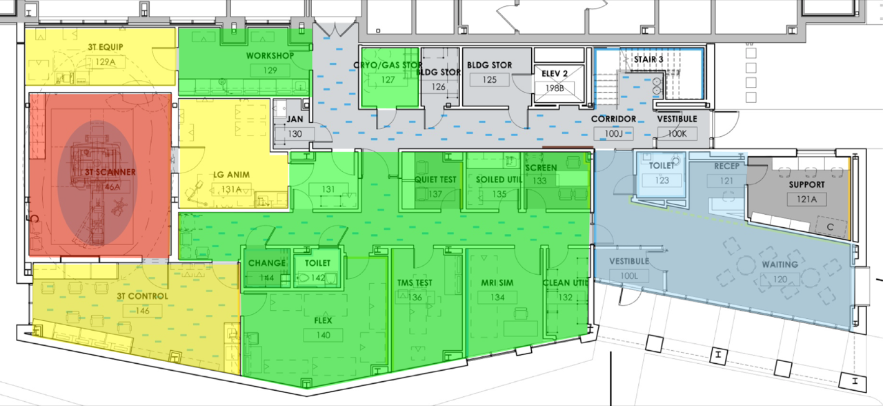
Zone I
Zone I consists of all areas freely accessible to the general public. This zone includes the entrance to the MR facility and the magnet poses no hazards in these areas.
Zone II
Zone II acts as a buffer between Zone I and the more restrictive Zone III. Here, patients are under the general supervision of MR personnel. Normally these areas are also safe from the powerful magnet. Zone II may include the reception area, dressing room, and interview room.
Zone III
Access to Zone III should be restricted by a physical barrier. Only approved MR personnel and patients that have undergone a medical questionnaire and interview are allowed inside Zone III. The MR control room and/or computer room are located within Zone III. Because the magnetic field is a three-dimensional volume, Zone III must be restricted and field hazards should be clearly delineated and restricted from non-MR personnel.
Zone IV
Zone IV is strictly the area within the walls of the MR scanner room, sometimes called the magnet room. Access into the MR scanner room should be inside of Zone III because it does not have a direct entrance point to unrestricted areas. Zones III and IV are sometimes collectively referred to as the MR suite.
Inside of the MR Suite is an invisible boundary defined by the magnetic field’s five Gauss line. The five Gauss line is the point at which the magnetic field begins to affect electromagnetic devices, such as pacemakers. Because the magnetic field extends in all directions, the five Gauss line can also extend to areas outside of the MR Suite, including other floors, if the magnetic field is large enough. Magnetic fields cannot be seen or felt, so the five Gauss line is sometimes marked on floors or walls of the MR scanner room.
Device and Object Screening
Any device entering Zone IV must have prior authorization from the center manager.
For informational purposes, the classification system of items that have the potential to come into contact with the magnetic field is below:

MR SAFE
MR Safe Objects pose no know hazards in the MR Environment.
They tend to me made of plastic, rubber, wood, or other non-magnetic, non- metallic, and non-conductive materials.
Importantly, there are certain patient care devices such as wheelchairs and gurneys that may be used safely in the MRI environment. Under no circumstances should you bring any outside gurneys or wheelchairs into the MRI lab area. Subjects should be moved to the safe devices while outside of the MRI console room.
MR Conditional
MR Conditional Objects pose no know hazards in the MR Environment under specific conditions such as field strength, position, etc.
MRI Conditional devices have been tested in the MRI environment under very specific conditions of use, including instrument field strength, location in the room, position on the body etc… Most MRI fire extinguishers are labeled “Conditional” reflecting the fact that caution is still required in their use.
MR UNSAFE
MR Unsafe objects are not to be brought into the MRI Environment under any circumstances.
Most devices should be assumed to be MR Unsafe, unless specifically labeled otherwise. This specific label usually is applied to objects that are intended to be used in an MRI facility but not in the magnet room. MR Unsafe labeling is relatively rare, and you should not assume that the lack of such labeling suggests that the devices are safe.
Trevor Wigal
Manager, CBBI
Phone: 302-831-1463
Xingju Nie
MRI Physicist
Phone: 302-831-0648
Ibrahim Malik
Research Associate
Phone: 302-831-0910
Sarah Whitman
Research Associate
Phone: 302-831-0910
HOURS OF OPERATION
The University of Delaware Research Office is open daily Monday through Friday, 8 a.m.– 5 p.m. The Research Office is closed Saturday and Sunday and observes all University closings and holidays. See the Academic Calendar for more information.
If you have an emergency during non-business hours, please call Public Safety at 302-831-2222.
If your question does not require immediate attention, please e-mail CBBI.