

Capabilities
CBBI is the home for neuroimaging research at the University of Delaware. The centerpieces of the facility are the state-of-the-art Siemens 3T Magnetom Prisma and Bruker Biospec 94/20 MRI scanners.

Our Siemens Magnetom Prisma whole-body MRI scanner, with its 3 Tesla magnet, provides stunningly detailed images, allowing researchers in multiple disciplines a safe, non-invasive way to study the structure and function of the human brain as well as the sructure of bone, joints, and muscle in children and adults.
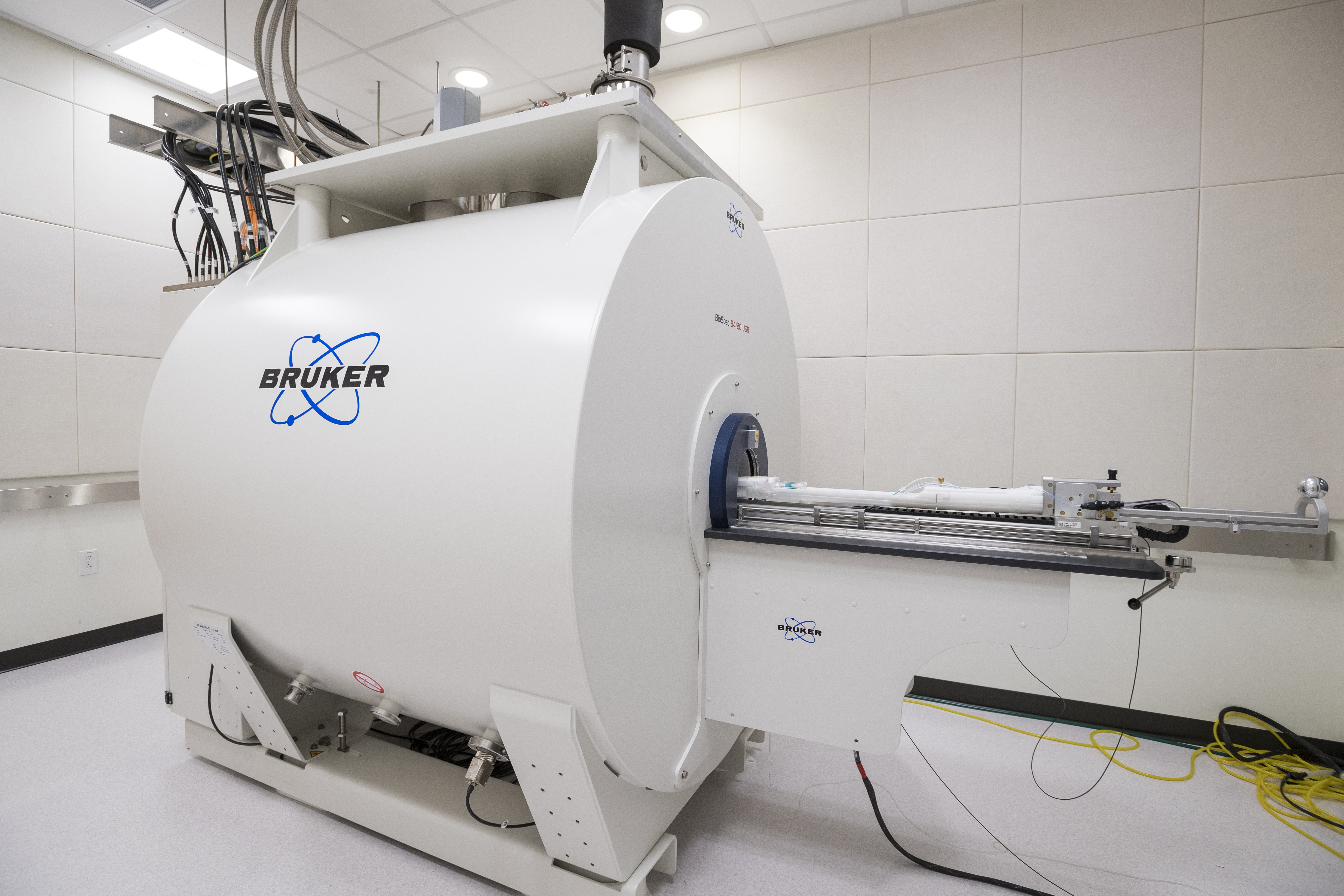
Our Bruker Biospec 94/20, a 9.4 Tesla magnet, designed for pre-clinical MR imaging and MRI research. State-of-the-art MRI technology combined with the ultra high field magnetic field delivers high spatial resolution, enabling researchers to come closer to the molecular and cellular level research they desire.
Details of the technical specifications and capabilities of the MRI scanners are provided below
Siemens 3T Magnetom Prisma MRI
Bruker Biospec 94/20 MRI
Outstanding gradient performance
- 80 mT/m @ 200 T/m/s simultaneously, on all three axes
- Ultra-high-performance cooling
- Force-compensated design for reduced vibrations
Benefits: Significantly increased SNR, unprecedented long-term stability and minimized acoustic noise.
Tim 4G integrated receive architecture
- New Tim 4G RF system with 48, 64 or 128 independent channels for faster imaging and higher SNR
- New Tim 4G coil technology with highest coil element density, Dual-Density Signal Transfer, DirectConnect, and SlideConnect technology
- Fully digital transmit and receive with DirectRF
- Real-time feedback loop for excellent long-term stability
Fully dynamic parallel transmit with TimTX TrueShape
- Selective excitation to highlight regions, organs, or even features of an organ
- Zoomed imaging with the first parallel transmit application syngo ZOOMit
- Significantly higher resolution, reduced scan times and less artifacts.
- Open platform for new MRI applications
Unmatched 3T magnet
- Based on the MAGNETOM Trio experience
- Benchmark magnet homogeneity
- FoV 50x50x50 cm3
- Advanced higher order shim and optional SpectroShim
- Zero helium boil-off
Ultra high-field MRI
- 9.4T static magnetic field
- Active shielding with reduced stray field
- Zero helium boil off
Benefits of Ultra High Field Imaging
- Increased SNR for higher resolutions and shorter scan times
- Increased Blood Oxygen Level Dependent (BOLD) contrast
- Higher magnetic susceptibility in fMRI, SWI, and QSM
- Increased chemical shift
- Increased spectral dispersion for spectroscopy and Chemical Exchange Saturation Transfer (CEST) Imaging
Other Features
- Paravision 360 operating suite
- TeamViewer remote support
- X-nuclei imaging
MAGNETOM Prisma offers:
Diffusion Spectrum Imaging
- Up to 514 diffusion directions
- Sensitive to multiple diffusion directions within a voxel
- Potential to characterize crossing fibers
TimTX TrueShape and syngo ZOOMit
- Finest imaging details from the most hidden structures
- High resolution DWI
- DTI Depicting structural CNS connectivity
Most powerful shimming system
- Benchmark magnet homogeneity
- Advanced higher order shim, optional SpectroShim
- Higher specificity in body and multinuclear spectroscopy
Excellent flow sensitivity and time-resolved Angiography with XR 80/200
- Depiction of smaller vessels without contrast with ToF
- Enhanced flow effects with XR 80/200 gradient system
- Higher SNR
- Extreme iPAT performance with Tim 4G
syngo DTI with up to 256 directions
- High-resolution isotropic DTI for finer depiction of white matter tracts
Most advanced tools in syngo.via
- Fully integrated fMRI/DTI evaluation
- Interactive track exploration mode with multiple VOI objects
Excellent long-term stability
- Excellent signal stability even for demanding long measurements – thanks to the new XR 80/200 gradient cooling system
And more…
- Cardiac Flow Imaging
- High-resolution joint imaging with excellent FatSat
- Zoomed high-resolution imaging of the pelvis
- High-resolution with isotropic 3D imaging
- Up to 2x more SNR in diffusion studies
- Advanced Flow Evaluation
- Advanced multi-nuclear Spectroscopy
Peripheral Support Equipment
Visual stimulation: BOLDscreen 32 LCD display; VPixx ProPixx projector for high temporal frequency (up to 1440 Hz) and dichoptic stimulation with DataPixx Lite data acquisition system; CinemaVision video goggles by Resonance Techbology; MediGlasses MR safe prescription glasses set
Auditory stimulation: MR Confon F MK II package (headphones); Sensimetrics ear buds; OptoAcoustics OptoACTIVE noise canceling headphones
Eyetracking: SR Research EyeLink 1000 Plus
Subject response devices: Current Designs fORP 932 8-channel response system with bimanual 8-button response box, 4-button response box and 2-button response box (´2); Tethyx joystick and trackball; Foot pedal; OptoAcoustics FOMRI-III MRI microphone; Black Box Toolkit v2 Elite–36 channels
Physiological monitoring: BIOPAC MP150 (ECG amplifier, respiration transducer, skin transducer); Siemens PMU (ECG, finger pulse and respiration); MRC in-bore video camera 12M-I with infrared illuminator
ASSISTANCE
Keith Schneider
Director, CBBI
Professor, Psychological & Brain Sciences
Phone: (302)-831-7148
Trevor Wigal
Manager, CBBI
Phone: 302-831-1463
Xingju Nie
MRI Physicist
Phone: 302-831-0648
Ibrahim Malik
Research Associate
Phone: 302-831-0910
Sarah Whitman
Research Associate
Phone: 302-831-0910
HOURS OF OPERATION
The University of Delaware Research Office is open daily Monday through Friday, 8 a.m.– 5 p.m. The Research Office is closed Saturday and Sunday and observes all University closings and holidays. See the Academic Calendar for more information.
If you have an emergency during non-business hours, please call Public Safety at 302-831-2222.
If your question does not require immediate attention, please e-mail CBBI.